 |

Search
|
|
|
Equine Mandibular Juvenile Ossifying Fibromas
Although equine tumors are fairly uncommon, a significant portion of those that do arise occur in the head and neck region. Specifically, tumors of the oral cavity may originate in the mandible, gums, tongue, etc., often extending into the surrounding tissues. One of these tumors is the ossifying fibroma that tends to develop from the intramembranous bone of the mandible. This tumor has a high occurrence in young (2-14 months of age) horses, no breed or sex predilection has been shown, and genetic predisposition has yet to be determined. Many believe this tumor can be diagnosed through history, signalment, physical examination and radiographic findings, as it presents with highly characteristic features. Nevertheless, in order to definitively diagnose an ossifying fibroma and distinguish it from similar proliferative lesions in the mandible (osteoma, osteosarcoma, fibrous dysplasia, fibrous osteodystrophy), histo-pathology should be key in the diagnostic plan.
This neoplasm is locally aggressive, with extensive bony proliferation and trabecular destruction, yet there are no reports of it possessing metastatic qualities. Medical and/or surgical options may be employed to successfully treat the neoplasm, avoiding further occurrence. However, as with other invasive neoplastic processes, inadequate resection of margins or incomplete treatment often leads to rapid, extensive and increased regrowth. Prognosis is often dependent on the extent of mandibular involvement (prehension difficulty) and aesthetic appearance of the horse (owner's visual value).
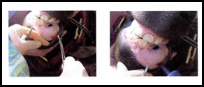
Clinical Presentation and History: Clinical signs associated with the juvenile ossifying fibroma depend on location and size of the tumor. They may include difficulty with prehension, lymphadenopathy, and intermittent oral mucosal bleeding. Early diagnosis is not often made because the anatomy of the oral cavity allows for a considerable amount of involvement and progression of the ossifying fibroma before obvious clinical signs are noted. When clinical signs are apparent, advanced local infiltration is often present. The most common presentation is as a sub-gingival, bony proliferation on the rostral mandible in a young horse. The mass is uniformly firm and does not elicit signs of pain when manipulated. The
mucosa covering the mass is usually ulcerated. Upon palpation, the teeth in the affected area may be loose.
It is controversial whether the juvenile mandibular ossifying fibroma is caused by trauma, or whether an undetected, immature fibroma causes the bone to be brittle and more susceptible to minor trauma. Many of the reported cases cite an injury as the cause of the bony proliferation. Trauma affecting the mandible through a fall, kick or self-inflicted injury (e.g., running into objects), will often result in gingival ulcerations and tears that will not heal despite weeks to months of treatment, progressing to the growth of a prominent hard structure at the site of injury. The ossifying fibroma arises from mutations in normal bony remodeling that would otherwise reconstruct the mandible. The mass will proliferate until the lips are no longer apposed (allowing visualization of the mass), prehension difficulty is noted, and weight loss occurs as a result of not eating. Many feel that the trauma sustained by the mandible should not be substantial enough to elicit such an injury with obvious prolonged healing time. Therefore, another facet to the trauma theory is that the ossifying fibroma already existed in the bone, had weakened its trabecular structure, and made the mandible more susceptible to minor injury. Extensive and rapid growth is then stimulated by the trauma. |
 |
Diagnosis: A tentative diagnosis can be made based on the history, as well as on the gross, clinical and radiographic aspects of the lesion.
Histological examination of the mass is used to confirm the presumptive diagnosis. |
This can be done on a core biopsy or an en bloc excisional biopsy. The juvenile mandibular ossifying fibroma is characterized by well differentiated, moderately vascularized, abundant, dense fibroblastic stroma, with isomorphic fibroblasts transforming into osteoblasts that rim bony spicules. The histologic alterations tend to be very uniform in appearance throughout the mass.
To distinguish the juvenile mandibular ossifying fibroma from other closely resembling non-neoplastic and neoplastic lesions, histologic morphology plays an important role. Unlike ossifying fibroma, bony spicules in fibrous dysplasia are rarely lined by osteoblasts, and only mature lesions contain deposits of lamellar bone. Another differential diagnosis can be osteoma. These are bony growths that are initially formed of cancellous bone with intertrabecular fatty or hematopoietic marrow; they may become increasingly compact with time. Because of the morphological similarity between ossifying fibroma and some cases of osteoma, it is thought that ossifying fibromas may mature into osteomas. Finally, in osteosarcomas, neoplastic cells have a high mitotic index and are pleomorphic, features which are lacking in ossifying fibromas.
Treatment: Treatment includes surgical (mandibulectomy, hemi-mandibulectomy) and medical (radiation therapy) management. Combinations of these therapies may also be employed.
Surgical management requires the extensive removal of the entire mass and involved structures (teeth), with achievement of adequate clean margins. It has been widely reported that local excision of a juvenile mandibular ossifying fibroma often results in rapid and proliferative recurrence unless the surgical excision includes wide surgical margins. The choice of which surgical procedure to use is based on diagnostic imaging (radiographs, computerized tomo-graphy), which determines the extent of bony involvement. If diagnosed or suspected early in the growth process, a rostral mandibulectomy or rostral hemi-mandibulectomy may suffice as proper treatment. If there is significant bony involvement, more drastic surgical procedures (complete mandibulectomy or hemi-mandibulectomy) are recommended. When the ossifying fibroma has grown from the rostral mandible, involved the entire mandibular symphysis, and extended to both hemi-mandibles, internal fixation (metal implants) must be used to create a pseudosymphysis upon removal of the neoplasm. This allows for proper apposition of dentition, as well as stabilization of the grinding forces of the jaw during mastication. If complete removal of the juvenile ossifying fibroma is achieved, there is a very low probability of recurrence, even years post surgery. If regrowth is to occur, most studies have shown that this takes place within the first six months post surgery.
Radiation therapy, the other therapeutic option in cases of mandibular ossifying fibromas, uses ionizing radiation to treat the neoplasm and to limit the neoplastic growth. While not surpassing the normal tissue tolerance of the healthy tissue surrounding the ossifying fibroma, radiation therapy delivers a sufficient lethal dose of radiation to the tumor tissues. Radiation therapy can be delivered through brachytherapy or, more commonly, through an external beam. External beam therapy includes gamma or X-rays from megavoltage equipment with Cobalt-60, linear accelerators or orthovoltage machines. Success has been obtained by treating the ossifying fibroma with a bilateral parallel opposed pair technique. The radiation margins should include the tumor and a border of normal, healthy tissue. After several successive treatments, the mass initially appears to be the same size, but less radiodense using diagnostic imaging. Over time, the ossifying fibroma progressively decreases in size to the point of no visible external existence.
As with radiation treatment, serial follow-up radiographs are extremely important in the surgical post-operative monitoring of the patient. Radiation therapy can be combined with surgery; surgery can be used to either debulk the mass for radiation therapy or used in en bloc excision to expose transitional margins primed for radiation therapy.
In summary, mandibular juvenile ossifying fibroma is a locally invasive, proliferative, fibro-osseous tumor that is most commonly found in the mandible of young horses. Though aggressive in nature, the neoplasm is benign, as no incidents of metastasis have been reported. Grossly it is very distinct, yet in order to definitively diagnose this mass, histopathology must be employed. If diagnosed prior to significant mandibular involvement, treatment options yield a fair to good prognosis. Both surgical and radiation therapies have resulted in extremely low recurrence rates when adequately employed, with the horse returning to normal prehension, activity and visual aesthetics post treatment.
-by Araba Oglesby, Class of 2006
-edited by Dr. Ingeborg Langohr
References
-
Bertone J and Brown CM: 2003. The 5-Minute Veterinary Consult-Equine. Lippincott, Williams and Wilkins. Iowa State University, December.
-
Collins JA: 1998. Ossifying fibroma/osteoma in the proximal tibia of a mature gelding. Vet Record 143(13): 367-368.
-
Hance SR, Bertone AL: 1993. Neoplasia. Vet Clin North Am Eq Pract. 9(1):213-234.
-
Morse CC, Saik JE, Richardson DW, Fetter AW: 1988. Equine juvenile mandibular ossifying fibroma. Vet Pathol 25(6): 415-21.
-
Orsini JA, Baird DK, Ruggles AJ: 2004. Radiotherapy of a recurrent ossifying fibroma in the paranasal sinus of a horse. J Am Vet Med Assoc 224(9): 1483-1486.
-
Richardson DW, Evans LH, Tulleners EP: 1991. Rostral mandibulectomy in five horses. J Am Vet Med Assoc. 199(9): 1179-1182.
-
Roberts MC, Groenendyk S, Kelly WR: 1978. Ameloblastic odontoma in a foal. Equine Vet J 10(2): 91-93.
-
Robbins SC, Arighi M, Ottewell G: 1996. The use of megavoltage radiation to treat juvenile mandibular ossifying fibroma in a horse. Can Vet J. 37(11): 683-684.
-
www.vin.com (search juvenile ossifying fibroma
|
|
|
|
|