FINAL DIAGNOSIS: This column in the Winter 2006 issue is being replaced by an article written by Carla Vega de la Cruz, Purdue Merck-Merial Summer Research Scholar, Tuskegee University
Bone Marrow Fat Analysis as a Measure of Starvation in Animals
Summary: Making a definitive diagnosis of starvation in animals is difficult because there are few quantitative measures of starvation available at postmortem examination. The Toxicology and Analytical Chemistry section of the Purdue ADDL is currently developing a method which will be used to relate severely decreased bone marrow fat to clinical starvation. This will be done by developing an analytical method for bone marrow fat analysis, establishing a database of values for the normal percentage of bone marrow fat in domestic animals, and relating severely decreased bone marrow fat to clinical starvation. At this time, we welcome inquiries regarding the submission of femurs for analysis.
Rationale and Significance: Malnutrition is a state in which a diet does not provide the optimal amount of nutrients. The long-term effect of inadequate intake of food is starvation. Inadequate food intake can be exacerbated by a physiological condition or disease state as well as by extreme environmental factors such as those that occur in winter. Malnutrition and starvation are a natural cause of death in wildlife. Management practices resulting in malnutrition and starvation can also occur in domestic livestock. When this occurs, there can be legal ramifications related to mismanagement and mistreatment. However, diagnostically, there are few quantitative measures of starvation available at post-mortem examination. This is why making the definitive diagnosis of starvation is many times difficult, especially if the cause and time of death are unknown (Ballard, 1995). Therefore, a need exists for a validated, quantitative analytical method which can be used to support a post-mortem diagnosis of starvation.
Literature Review: Malnutrition is defined as the inadequate intake and/or malabsorption of any required nutrients (Stedman's, 1995). This can occur in an animal which is eating, but is not able to ingest, digest, absorb, and/or utilize a sufficient quantity of nutrients (Radostits, 2000). In addition to simple lack of food/nutritional intake, malnutrition can be related to injuries, bad teeth, parasitism, neoplasia, toxins, or infectious disease (Hungerford, 1990). Starvation is characterized by a lengthy and continuous deprivation of food (Stedman's, 1995). They both can be caused by diseases, injuries, management conditions, and/or the environmental conditions in which the animals live. In the northern hemisphere, winter can bring on additional stress to outdoor livestock due to a lack of food-related negative energy balance brought about by poor quality/inadequate forages, cold weather, and increased energy demands (Radostits, 2000).
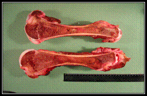
In wild ruminants such as deer and moose, analysis of bone marrow fat content by various methods has been used for several decades for diagnosis of starvation because, following harsh winters, bones are frequently the only sample which can be found for evaluation (Cheatum, 1949, Bischoff, 1954, Greer, 1968, Neiland, 1970, Verme and Holland, 1973, Franzmann and Arneson, 1976). In those studies, a fat solvent extraction method was generally found to provide the most consistent results when compared to other methods even though some of the other air-drying or compression methods are more rapid and easier to perform in the field (Greer, 1968, Meiland, 1970, Verme and Holland, 1973). In wildlife, the femur has been used as a standard when evaluating bone marrow fat content (Ballard, 1995). The femur is used because it is readily obtained, has a large marrow content, an abundant blood supply, and is one of the last fat sources to be utilized. The bone marrow of a normal healthy animal is solid, white and waxy due to the high fat content (Cheatum, 1949). In a state of malnutrition, the bone marrow is red, solid, and slightly fatty to the touch (Cheatum, 1949). In an advanced state of starvation, the bone marrow is red to yellow, gelatinous, and glistening and wet to the touch due to the high water content (Cheatum, 1949). In addition to the applicability of the solvent extraction method, findings of those wildlife studies pertinent to domestic livestock include: 1) with a high degree of accuracy, a gelatinous bone marrow ,regardless of its color, is indicative of a poor animal resulting directly or indirectly from malnutrition as in one study, 95% of gelatinous marrows were found in poor deer and 97% were low in marrow fat (low defined in that study as less than 19%, Bischoff, 1954), 2) no definite conclusion can be made from a solid marrow concerning deer condition as in some cases they can appear solid down to approximately 40%-50% fat (Bischoff, 1954), 3) tibia marrow does not correspond to femur marrow (Bischoff, 1954), 4) in another study, femur bone marrow fat content (by solvent extraction) in winter-killed elk was less than 0.25% (n=12) although the fat content in other live elk at the end of winter could be as low as 1% (Greer, 1968), and 5) in an additional study, femur bone marrow fat from winter-killed moose was as low as 6.1% fat in calves and 5.5% in adults by a dry-weight method which includes non-fat residue (Franzman and Arneson, 1976). However, while these studies have been performed on wildlife, there are no published reports of the use of bone marrow fat for diagnosis of starvation in domestic livestock.
The body utilizes different sources (carbohydrates, protein and fat) for energy. Generally, the first source utilized is carbohydrate in the form of glycogen. However, glycogen stores are relatively rapidly exhausted and the next source for energy is predominantly fat. Bone marrow fat is one of the last body stores of fat to be used. Late in the course of starvation, when glycogen and fat stores have been depleted, the only source available for energy is protein, the catabolism of which results in the development of ketosis (ketone bodies in blood and urine). If this negative energy balance is not corrected the animal will die. In general, clinical signs and gross pathological findings related to malnutrition/starvation include animals that are weak and underweight, have a loss of skin turgor, have dull hair coats, sunken eyes, tucked-up abdomens, prominence of the bones of shoulders, ribs, vertebra and pelvis, atrophy of muscles, and a decrease or absence of subcutaneous, perirenal, pericardial and bone marrow fat which can be described as serous atrophy of fat.
|