Granulomatous meningoencephalomyelitis (GME) in dogs
GME is an acute, progressive inflammatory disease of the central nervous system (CNS) of dogs. GME is a common differential for dogs that are affected by focal or diffuse neurological diseases. An inflammatory disease like GME can cause severe and often irreversible damage to the CNS. Consequently, a better understanding of the disease is essential.
Etiology: GME has been reported around the world and can affect most breeds and ages of dogs; however, middle aged, small breed dogs such as terriers and poodles are more susceptible (Thomas, 1998). GME accounts for up to 25% of all canine CNS disorders reported in the United States (Cuddon, 1984). No specific etiological agent has been described for this disease.
Clinical signs: The clinical signs of the disease are variable depending on the location of the lesion in the CNS. Three syndromes of GME have been recognized based on the location of the lesion:
a) Focal GME - this is a chronic progressive condition (3-6 months) and the clinical signs occur secondary to nodular granuloma formation and mimic the effects of space occupying tumor/masses.
b) Multifocal or disseminated GME - This is an acute, progressive condition (2-6 weeks). The most common sites affected are lower brain stem, cervical spinal cord and meninges. Up to 25% of the dogs are dead within a week (Wong and Sutton, 1002).
c) Ocular form- this can be acute, progressive or static and can affect eyes unilaterally or bilaterally.
Depending on the location of the lesions, the clinical signs can vary, but neurological deficits and pain from meningeal involvement are common.
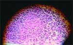 Pathology: At necropsy, gross lesions are evident if the angiocentric inflammation is severe and can be seen as areas of swelling and yellow to gray discoloration. Histopathologic lesions are characterized by perivascular cuffs of monocytes, macro-phages, lymphocytes and plasma cells. These perivascular cuffs merge at adjacent blood vessels to form cellular whorls that can evolve into nodular granulomas (Ryan et al (Ryan et al, 2001). Immunohistochemical characterization of the inflammatory cells in the granulomatous lesions of GME showed that the lesions consist of MHC class II and CD3+ T-cells indicating a T-cell mediated delayed hypersensitivity reaction (Kipar, 1998).
Diagnosis: GME diagnosis is supported by the exclusion of neoplastic, infectious and other inflammatory conditions (e.g., canine necrotizing meningoencephalitis, NME). CT and MRI can sometimes be of use in detection of the CNS lesions but it is difficult to differentiate the lesions from neoplasia. Cell characteristics such as cytologic atypia and mitotic figures might be useful to differentiate this condition from neoplasia or neoplastic reticulosis. Granulomatous inflammation due to viruses (e.g., rabies or canine distemper), protozoa (e.g., Toxoplasma and Neosporum), and fungi (e.g., Cryptococcus) can be ruled out by demonstration of specific antigens in CSF or serum antibody titers for the various etiologic agents. NME can be differentiated based on breed predilection (small-size breeds, especially Pugs) and lack of obvious granulomas.
Treatment: B) Corticosteroids are the mainstay of treatment for GME. Response to therapy is variable and discontinuation results in recurrence of clinical signs and progression of the disease. B) Leflunomide a de novo pyrimidine synthesis inhibitor can also be used because of the immune component (T-cell mediated) in the disease. However, these drugs are expensive and controlled clinical trial results are not available. C) Radiation therapy can prolong the mean survival (MST) of dogs.
Prognosis: The prognosis is generally poor for GME. MST for all dogs with GME is 14 days (range 1-1215 days - Munana, 1998). Dogs with focal signs in forebrain have an MST of >359 days while dogs with focal signs elsewhere have an MST of 59 days. Dogs with multifocal signs have an MST of 8 days. Dogs that receive radiation therapy for focal signs can survive >404 days. Corticosteroid therapy may induce a transient remission of clinical signs and can prolong the MST.
-by Jeetendra Eswaraka, EVFVG Student
-edited by Dr. Vimala Vemireddi, ADDL Graduate Student
References:
-
Cuddon PA and Smith-Maxie L: 1984. Reticulosis of the central nervous system in the dog. Comp Cont Educ Pract Vet 6:23-32.
-
Kipar A, Baumgartner W, Vogl C, Gaedke K and Wellman M: 1998. Immunohistochemical characterization of inflammatory cells in brains of dogs with granulomatous encephalomyelitis. Vet Pathol 35: 43-52.
-
Munana K and Lutgen P: 1998. Prognostic factors for dogs with granulomatous meningoencephalomyelitis: 42 cases (1982-1996). JAVMA 212: 1902-1906.
-
Ryan K, Marks SL and Kerwin SC: 2001. Granulomatous meningoencephalomyelitis in dogs. Compend Contin Educ Pract Vet 23(7): 644-650.
-
Thomas JB: 1998. Inflammatory diseases of the central nervous system in dogs. Clin Tech Small Anim Pract 13: 167.
-
Wong CW and Sutton RH: 2002. Granulomatous meningoencephalomyelitis in dogs. Aust Vet Pract 32(1): 6-11.