Hepatozoonosis is an emerging disease in
the United States caused by the protozoan Hepatozoon. Hepatozoon
species have been found to infect a wide range of carnivorous
hosts including domestic dogs, jackals, coyotes, foxes, hyenas,
domestic cats, bobcats, lions, leopards, and cheetahs. The
causative agent of hepatozoonosis in the United States is
a newly recognized species, Hepatozoon americanum, whereas
in other parts of the world, Hepatozoon canis is the primary
agent. The features of American hepatozoonosis greatly contrast
non-American infections. The clinical presentation of dogs
with hepatozoonosis in the US is much more aggressive than
that of infected dogs from other parts of the world, indicating
that Hepatozoon americanum is more pathogenic. Non-American
infections are often subclinical and seem to be limited to
the immunosuppressed. Most cases of domestic canine hepatozoonosis
in the United States are diagnosed in the region from Texas
to Georgia. H. americanum infection has also been identified
in coyotes in Oklahoma.
The exact life cycle and transmission for H. americanumhas
not been completely elucidated. Most portrayals are based
on extrapolations from that which is known of H. canis. The
tick vector is the definitive host. The vector for H. canis
appears to be primarily the brown dog tick, Rhipicephalus
sanguineus. Evidence suggests the Gulf Coast tick, Amblyomma
maculatum, as the vector involved in transmission of H.americanum
in the United States. Although some reports have demonstrated
the ability of dogs to become infected with H. americanum
after ingesting an infected R. sanguineus transmission from
an infected dog to the tick has not been documented.
The tick becomes infected by ingesting a blood meal, containing
monocytes or neutrophils laden with isogamonts from an infected
vertebrate host. Syngamy occurs in the gut of the tick, producing
a zygote which penetrates the tick gut wall. Sporogony occurs
in the haemocoel where an oocyst is formed containing multiple
sporozoites. The intermediate vertebrate host must ingest
the tick to become infected since the sporozoites apparently
do not migrate to salivary tissue in the tick. Sporozoites
penetrate the intestinal wall of the intermediate host and
undergo schizogony, forming schizonts, and then cysts within
mononuclear phagocyte or endothelial cells of the spleen,
bone marrow, lungs, liver, lymph node, or muscle. When the
schizonts rupture, an inflammatory reaction is initiated.
In completion of the life cycle, gamonts are produced that
infect circulating leukocytes. A paratenic prey host in which
only cysts are formed has not been documented with H. americanum,
and the feeding of encysted meat to carnivorous hosts has
not produced infection; however, such paratenic hosts have
been identified with otherHepatozoon species.
Immunosuppression seems to be an important determinant
of susceptibility for infection with Hepatozoon species.
Concurrent infection, debilitating disease, immunosuppressant
drugs, and young age seem to influence clinical manifestation.
It is unclear if clinical hepatozoonosis occurs only in the
immunosuppressed, or if infection elicits immunosuppression
in the host, predisposing to concurrent infection.
American hepatozoonosis typically presents as severe clinical
disease. A majority of the clinical syndrome is composed
of clinical signs related to chronic inflammatory disease.
Many patients present with recurrent fever, lethargy, depression,
and weight loss. Muscular disease is also apparent on presentation.
Schizogony of the hepatozoon causes a marked pyogranulomatous
polymyositis which results in stiffness, lameness, hyperesthesia,
and muscle atrophy. Clinical signs fail to resolve with antibiotics.
Bloody diarrhea, related to intestinal penetration by the
sporozoites, may be documented soon after exposure. A generalized
lymphadenomegaly may also be present.
Clinical pathological changes are often typified by a
marked, mature neutrophilic leukocytosis. A mild nonregenerative
normocytic, normochromic anemia is often observed. An eosinophilia
or thrombocytosis may be evident. Hypoglycemia, low urea
nitrogen, hypoalbuminemia, and hyperglobulinemia are other
common findings.
Electromyography may indicate a generalized polymyopathy.
Radiography of the appendicular skeleton may reveal disseminated
periosteal bone proliferation mostly involving the diaphysis
of long bones. Histopathological examination of the osseous
lesions displays changes that closely resemble those of hypertrophic
osteopathy. New spicules of bone forming in the periosteum
are oriented perpendicular to the cortex without producing
cortical destruction. The pathogenesis of such changes is
not well understood. Changes do not seem to be associated
with the presence of parasites or inflammation in the adjacent
skeletal muscle.
Immune-complex hypersensitivity resulting in fatal vasculitis
or glomerulonephritis is a potential sequela of infection.
A protein-losing nephropathy may be diagnosed in dogs with
glomerulonephritis.
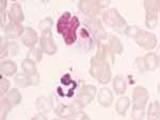
Definitive diagnosis is made by microscopic observation
of the organism. Gamonts may be detected in neutrophils and
monocytes on peripheral blood smears, although this finding
is more typical of Hepatozoon canis infection. Successful
diagnosis is usually achieved via muscle biopsy where developing
organisms are usually abundant. Histopathological changes
often consist of pyogranulomatous myositis, muscular necrosis,
and muscular atrophy. Hepatozoon cysts, or zoites contained
within acute granulomas, may be identified interspersed between
the muscle fibers. Lesions similar to those found in skeletal
muscle may also be found in cardiac muscle and smooth muscle
of the intestine. Lymph node aspirates often yield reactive
lymphoid hyperplasia, but organisms are very rarely found.
Bone marrow aspirates may indicate granulocytic hyperplasia
and erythroid hypoplasia, and also rarely yield organisms.
Pyogranulomas and H. amer-icanum cysts may also be
found in pancreas, lymph nodes, kidney, spleen, and lung.
Since Hepatozoon americanum causes a relatively
low level of parasitemia in comparison to H. canis,
antibody production rate may be lower than expected and therefore
cause higher numbers of false negatives when tested serologically.
Studies have not been performed to identify the prevalence
of uninfected or subclinically infected seropositive dogs.
Future studies are needed to evaluate the clinical efficacy
of serological diagnosis.
-by Michelle Dennis, Class of 2002
-edited by Dr. Mika Tanabe, ADDL Instructor
References
Craig TM, Green CE: 1998. Hepatozoonosis. Infectious Diseases
of the Dog and Cat, 2nd ed.: 458-465.
Kocan AA, Cummings CA, Panciera RJ, Mathrew JS, Ewing,
SA, Barker RW: 2000. Naturally occurring and experimentally
transmitted Hepatozoon americanum in coyotes from Oklahoma.
Journal of Wildlife Diseases 36(1): 149-153.
Macintire CK, Vincent-Johnson N, Dillon AR, Blagburn B,
Lindsay D, Whitley EM, Banfield C: 1997. Hepatozoonosis in
dogs: 22 cases (1989-1994). JAVMA 210(7): 916-22.
Matthew JS, Ewing SA, Panciera RJ, Woods JP: 1998. Experimental
transmission of Hepatozoon americanum Vincent-Johnson et al,
1997 to dogs by the Gulf Coast tick,Amblyomma maculatum Koch.
Veterinary Parasitology 80 (1) 1-14.
Panciera RJ, Mathew JS, Ewing SA, Cummings CA, Drost WT,
Kocan AA: 2000. Skeletal lesions of canine hepatozoonosis
caused by Hepatozoon americanum. Veterinary Pathology 37(3):225-230.
Panciera RJ, Ewing SA, Mathew JS, Lehenbauer TW, Cummings
CA, Woods JP: 1999. Canine hepatozoonosis: comparison of lesions
and parasites in skeletal muscle of dogs experimentally or
naturally infected with Hepatozoon americanum. Veterinary
Parasitology 82(4): 261-272.
Vincent-Johnson NA, Macintire DK, Lindsay DS, Lenz SD, Baneth
G, Shkap V, Blagburn BL: 1997. A new Hepatozoon species from
dogs: description of the causative agent of canine hepatozoonosis
in North America. Journal of Parasitology 83(6): 1165-1172.
|